
Currently Empty: £0.00

Call Us:
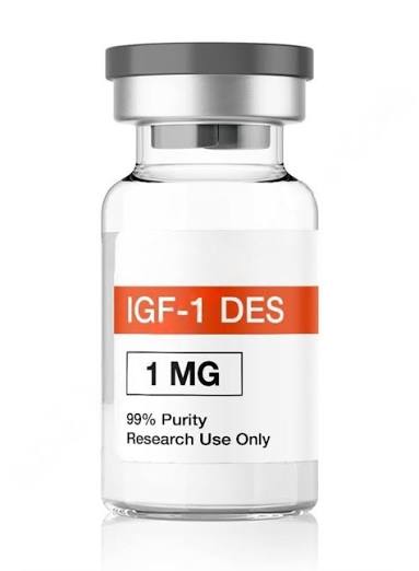


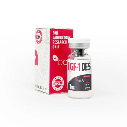
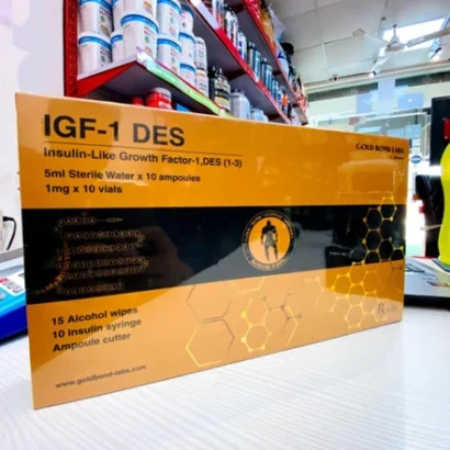
IGF-1 DES a remarkable example of how minor structural modification can produce profound pharmacological consequences. The proteolytic removal of just three N-terminal amino acids transforms a tightly regulated endocrine hormone into a potent, locally acting growth factor with approximately 10-fold greater biological activity than its parent molecule.
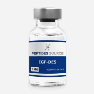
Price range: £45.00 through £715.00
IGF-1 DES is the primary mediator of growth hormone’s anabolic effects in peripheral tissues. When growth hormone is released from the pituitary and reaches the liver, it stimulates the production and secretion of IGF-1 DES into the bloodstream. This endocrine IGF-1 DES then acts on tissues throughout the body, driving cell proliferation, protein synthesis, and metabolic regulation through the IGF-1 receptor (IGF-1R). However, the IGF-1 DES system is far more complex than a single peptide—the IGF-1 gene undergoes alternative splicing to produce multiple variants with different tissue distributions, binding protein affinities, and biological activities.

Among these variants, IGF-1 DES—formally designated Des(1-3) IGF-1 or destripeptide IGF-1—stands out as one of the most potent and pharmacologically unique isoforms characterized to date. This naturally occurring truncated variant of human IGF-1, in which the first three N-terminal amino acids (glycine, proline, and glutamic acid) have been proteolytically removed, was first isolated from human fetal brain tissue in 1986.
The resulting 67-amino-acid peptide (molecular weight approximately 7,365 Da) has since been identified in bovine colostrum, porcine uterus, and various other tissues, establishing it as an endogenous bioactive IGF-1 isoform rather than a purely synthetic construct.
This production provides a comprehensive examination of IGF-1 DES, covering its molecular structure, mechanism of action, pharmacokinetic properties, research applications, safety profile, and regulatory status.
The discovery of IGF-1 DES represents a significant milestone in understanding the complexity of the IGF system. In the 1980s, researchers identified a brain-specific variant of IGF-1 that lacked the first three amino acids and demonstrated greater biological potency than the full-length molecule. Sara VR, Carlsson-Skwirut C, Andersson C, and colleagues (1986) published the first identification of this truncated form of IGF-1 in human brain tissue extracts, demonstrating that this naturally occurring variant was the predominant form of IGF-1 in brain tissue.
Subsequent research by Ballard FJ, Francis GL, Ross M, and colleagues (1987) provided systematic characterization of Des(1-3) IGF-1, showing approximately 10-fold greater potency than native IGF-1 in stimulating cell proliferation in vitro. This work established that the enhanced potency was attributable to reduced binding to IGF-binding proteins (IGFBPs) rather than increased receptor affinity.
The variant was also independently isolated from bovine colostrum, where it represents approximately 1-4% of total IGF-1 immunoreactivity, suggesting a physiological role in neonatal gut development. Research with the L6 myoblast cell line further elucidated the enhanced potency of this growth factor, providing a cellular model for understanding its mechanism of action.
3.1 Primary Structure
IGF-1 DES is a truncated form of human IGF-1 consisting of 67 amino acids. The parent molecule, full-length IGF-1, is a 70-amino-acid single-chain polypeptide. IGF-1 DES lacks the first three N-terminal residues—glycine (Gly), proline (Pro), and glutamate (Glu)—yielding a molecular weight of approximately 7371.4 Da compared to 7649 Da for intact IGF-1. The amino acid sequence of IGF-1 DES is:
TLCGAELVDALQFVCGDRGFYFNKPTGYGSSSRRAPQTGIVDECCFRSCDLRRLEMYCAPLKPAKSA
The CAS registry number for IGF-1 DES is 112603-35-7. Its molecular formula is C₃₁₉H₄₉₅N₉₁O₉₆S₇.
3.2 Three-Dimensional Structure
The truncation preserves the overall three-dimensional fold of IGF-1, which adopts a compact structure stabilized by three intramolecular disulfide bridges: Cys6-Cys48, Cys18-Cys61, and Cys47-Cys52 (numbered relative to the full-length sequence).
These disulfide bonds organize the peptide into a characteristic insulin-family fold consisting of three alpha-helical segments connected by loop regions. The B domain, C domain, and A domain architecture that IGF-1 shares with insulin and IGF-2 remains intact in the DES variant, as the excised tripeptide resides upstream of the first structured helix.
3.3 Structural Basis of Functional Modification
The removal of the Gly-Pro-Glu tripeptide from the N-terminus represents a structurally conservative but functionally transformative modification. Structural studies of full-length IGF-1 bound to IGFBPs have demonstrated that the N-terminal residues form critical contacts with the hydrophobic binding cleft of IGFBP-3 and other family members. Specifically, the Glu3 side chain participates in electrostatic interactions and hydrogen bonding with IGFBP residues that stabilize the IGF-1/IGFBP complex.
The loss of these three residues reduces IGFBP-3 binding affinity by approximately 100-fold. This reduction extends across the family of six high-affinity IGFBPs (IGFBP-1 through IGFBP-6), though the magnitude of binding reduction varies among individual members. The consequence is that IGF-1 DES exists predominantly in its free, unbound state in biological systems, rather than sequestered in the ternary complex of IGF-1/IGFBP-3/acid-labile subunit (ALS) that normally carries more than 99% of circulating IGF-1.
Importantly, the N-terminal truncation does not significantly alter the affinity of IGF-1 DES for the IGF-1 receptor (IGF-1R). The receptor-binding determinants of IGF-1 are located primarily in the B and A domains, particularly residues in helical segments that contact the L1 and CRD domains of the IGF-1R extracellular region. Because these determinants are preserved, IGF-1 DES binds to the receptor with affinity comparable to that of native IGF-1.
4.1 Binding Protein Evasion
The fundamental mechanism underlying IGF-1 DES’s enhanced potency is its dramatically reduced affinity for IGF-binding proteins. Over 95% of circulating full-length IGF-1 is sequestered by IGFBPs, which serve as a reservoir that extends IGF-1’s circulating half-life from minutes (free IGF-1) to approximately 12-16 hours (bound in the ternary complex). IGFBPs also regulate IGF-1 bioavailability—only free IGF-1 can bind to the IGF-1R and activate downstream signaling.
The N-terminal tripeptide is critical for binding to IGFBPs, particularly IGFBP-3 and IGFBP-5. With this region deleted, IGF-1 DES exhibits up to 100-fold reduced affinity for IGFBPs, meaning it circulates and acts locally in a predominantly free, unbound state rather than being sequestered in the IGFBP-3/ALS ternary complex that normally regulates native IGF-1 bioavailability.
4.2 Receptor Activation
Despite the N-terminal truncation, IGF-1 DES retains full binding affinity for the IGF-1 receptor, as the receptor-binding domain resides in the central and C-terminal regions of the peptide. In fact, the N-terminal modification paradoxically enhances binding affinity for the IGF-1 receptor by approximately 10-fold compared to native IGF-1, likely through improved access of the modified N-terminus to the receptor binding cleft.
The dramatically increased free fraction means that at equivalent total concentrations, IGF-1 DES produces substantially greater IGF-1R occupancy and activation than native IGF-1. The combined effect is a molecule with approximately 10 times greater biological potency than native IGF-1 per unit free concentration, compounded by the absence of IGFBP-mediated sequestration. In biological systems where IGFBPs are abundant (which includes virtually all in vivo contexts), the effective potency differential can be substantially greater.
4.3 Downstream Signaling Pathways
IGF-1 DES activates two primary intracellular signaling cascades:
PI3K/Akt/mTOR Pathway: This pathway drives enhanced protein synthesis, ribosomal biogenesis, and anti-apoptotic signaling. The mTOR-mediated effects on p70S6K and 4E-BP1 phosphorylation are particularly potent, making IGF-1 DES a substantially more effective stimulator of muscle protein synthesis and hypertrophy. The IGF-1/PI3K/Akt pathway also prevents expression of muscle atrophy-induced ubiquitin ligases by inhibiting FOXO transcription factors.
Ras/MAPK/ERK Pathway: This pathway promotes cell proliferation and differentiation.
The enhanced potency of IGF-1 DES makes it approximately 10 times more mitogenic than native IGF-1 in cell culture studies. When administered locally (e.g., intramuscularly), it acts with rapid onset due to its free bioavailability, strongly activating satellite cell proliferation and differentiation—the muscle stem cells responsible for repair and growth.
4.4 Differential Tissue Effects
Research has demonstrated that administration of des IGF-1 has differential effects on the activation of the MAP kinase and PI 3-kinase pathways in mouse skeletal and cardiac muscle. Elevated IGF-1 has a hypertrophic effect on skeletal muscle only in growth situations, and transgenic mice that overexpress Class 2 IGF-1 Ea in skeletal myofibres show striking muscle hypertrophy.
5.1 In Vitro Potency
IGF-1 DES is approximately 10-fold more potent than native IGF-1 at stimulating hypertrophy and proliferation of cultured cells. This enhanced potency has been demonstrated across multiple cell types:
· Cell proliferation assays: IGF-1 DES was approximately 10x more potent than native IGF-1
· Protein synthesis in L6 myoblasts: Demonstrated dose-dependent stimulation at concentrations approximately 10-fold lower than required for equivalent stimulation by native IGF-1
· Muscle cell differentiation: Cells treated with des-IGF-1 demonstrated a 19-20 fold increase in creatine kinase (CK) activity versus untreated cells
· Motoneuron survival: Des(1-3) IGF-1 demonstrated 2.5 times greater potency than IGF-I in promoting motoneuron survival
5.2 IGFBP Binding Affinity
IGFBP binding affinity for IGF-1 DES is reduced approximately 100-fold compared to native IGF-1. This dramatic reduction in binding protein affinity is the primary mechanism underlying its enhanced bioavailability and potency.
5.3 IGF-1R Binding Affinity
IGF-1 DES binds to the IGF-1 receptor with affinity comparable to that of native IGF-1. Some studies suggest an approximately 10-fold higher binding affinity than native IGF-1. The binding is specific, as it can be totally displaced by increasing concentrations of unlabeled rh des(1-3) IGF-I.
5.4 Comparative Potency
In comparative studies, IGF-1 DES was 160-fold more potent than IGF-II in type 1 receptor-mediated assays, underscoring that IGF-I and IGF-II analogs are not interchangeable for type 1 receptor-mediated readouts.
6.1 Half-Life
IGF-1 DES has a characteristically short half-life, a direct consequence of its lack of IGFBP-mediated protection from proteolytic degradation.
· Serum half-life in rats: 20.5 minutes, compared to 228.3 minutes for native IGF-I
· Estimated half-life: Generally reported as 20-30 minutes
· Circulation time: 6-12 hours in circulation
· Full-length IGF-1 in ternary complex: Approximately 12-16 hours
· Free IGF-1: Approximately 10 minutes
The short half-life favors brief, localized signaling rather than sustained systemic exposure. This makes IGF-1 DES particularly suited for targeted tissue growth effects at the site of administration rather than systemic anabolic signaling.
6.2 Plasma Clearance
Radiolabeled des-(1-3)-IGF-1 exhibits a mean total plasma clearance rate of 4.59 mL/min/kg in rats, compared to 1.20 mL/min/kg for IGF-I and 1.34 mL/min/kg for IGF-II. Measurement of total plasma clearances shows the rapid removal of des(1-3)IGF-I compared with IGF-I and IGF-II. The mean steady-state volume of distribution is larger for des(1-3)IGF-I than for IGF-I and IGF-II.
6.3 Tissue Distribution
Maximal accumulation of all IGF peptides is found in the kidney. The distribution pattern for 125I-IGF-I and -II shows levels higher in kidney > pancreas > small intestine > liver > duodenum > stomach > lung > spleen > heart > large intestine > testis > brain > skeletal muscle. Skeletal muscle, kidney, and testis show preferential uptake of the variants. Urinary excretion of the variants is much greater, consistent with their more rapid clearance from circulation.
6.4 Blood-Brain Barrier Interactions
Des(1-3)IGF-1, an IGF-1 analogue with little protein binding but similar biological activity, has a shorter half-life in blood, slower influx rate into brain, and no alteration in pharmacokinetics after addition of nonradiolabeled peptide.
7.1 Cell Culture Applications
IGF-1 DES is widely used in cell culture research due to its enhanced potency and reduced IGFBP interference. Its applications include:
· Myoblast studies: Used to study muscle cell proliferation, differentiation, and hypertrophy
· Neuronal research: Supports neuronal expression and survival in organ culture models
· Cancer research: Used to study IGF-1R signaling in various cancer cell lines
· Stem cell research: Activates satellite cell proliferation and differentiation
7.2 Preclinical Research
IGF-1 DES has been investigated in various preclinical models:
· Muscle hypertrophy: Studies in mouse skeletal and cardiac muscle examining differential effects on ERKs, AKT-1, and P70 S6K activation
· Neuronal protection: Investigated for effects on neuronal loss after hypoxic-ischemic brain injury
· Olfactory bulb organ culture: Potently enhances differentiated cell growth
· Gut tissue: Selective anabolic effects particularly evident in gut tissues
7.3 Potential Therapeutic Applications
While IGF-1 DES has no approved therapeutic use, clinical opportunities have been considered for:
· Catabolic states: Conditions characterized by muscle wasting
· Inflammatory bowel diseases: Due to selective anabolic effects in gut tissues
· Neurological conditions: Due to its presence in brain tissue and neuroprotective properties
8.1 IGF-1 DES vs. IGF-1 LR3
IGF-1 LR3 (Long R3 IGF-1) is a synthetic analog containing 83 amino acids with an Arg substitution at position 3 and a 13-amino acid N-terminal extension. Key differences include:
Parameter IGF-1 DES IGF-1 LR3
Half-life 20-30 minutes 20-30 hours
Origin Naturally occurring Synthetic
Potency ~10x native IGF-1 ~3x native IGF-1
Action Localized, short-duration Systemic, long-acting
Dosing More frequent (1-2x daily) Less frequent
IGF-1 DES is described as “a highly concentrated lightning strike” compared to IGF-1 LR3 as “a systemic, long-acting wave”. IGF-1 DES is preferred for studying rapid, localized tissue-specific growth effects without prolonged systemic exposure.
8.2 IGF-1 DES vs. MGF
Mechano-Growth Factor (MGF) is a splice variant of IGF-1 produced in response to mechanical stimulation. Unlike IGF-1 DES, which is a truncated form of mature IGF-1, MGF contains a unique E-domain sequence and is involved in local tissue repair and regeneration.
9.1 General Safety Considerations
The safety profile of IGF-1 DES in humans has not been established. No controlled clinical trials, formal toxicology studies, or systematic adverse event monitoring have been conducted for this peptide. The information available is derived from preclinical observations, extrapolation from the known pharmacology of the IGF-1 signaling axis, and the clinical safety database of the related FDA-approved product mecasermin (recombinant human IGF-1). All side effects, contraindications, and interactions described should be considered theoretical or inferred unless otherwise noted.
9.2 Hypoglycemia
The most significant predicted adverse effect of IGF-1 DES is hypoglycemia. The IGF-1 receptor and the insulin receptor share substantial structural homology, and IGF-1 exhibits cross-reactivity with the insulin receptor at approximately 1/100th the affinity of insulin. However, because IGF-1 DES circulates in an almost entirely free (unbound) state due to its dramatically reduced IGFBP binding, the effective concentration of active peptide reaching insulin receptors is far higher than with equivalent doses of native IGF-1.
Clinical experience with mecasermin (Increlex) demonstrates that even protein-bound native IGF-1 causes clinically significant hypoglycemia, which is the most common serious adverse effect in treated patients. The prescribing information for mecasermin includes a boxed warning regarding hypoglycemia, with reported rates of symptomatic hypoglycemia in approximately 40-50% of treated patients during clinical trials. IGF-1 DES, with its approximately 10-fold greater bioavailability, would be expected to carry an even higher risk.
IGF-1 DES can activate insulin receptors at high local concentrations, causing potentially dangerous drops in blood glucose. The risk is amplified by the absence of IGFBP buffering, allowing rapid and unregulated receptor activation. Hypoglycemia is reported in 10-30% of cases.
9.3 Localized Tissue Growth
Potent local mitogenic and hypertrophic effects may cause disproportionate tissue growth at or near the administration site, particularly in muscle or connective tissue. This is reported in 1-10% of cases.
9.4 Injection Site Reactions
Injection site pain, redness, or swelling at subcutaneous or intramuscular injection sites occurs in 10-30% of cases. More broadly, localized injection site reactions (swelling, redness, tingling) affect 40-60% of users but typically resolve within 24-48 hours.
9.5 Cancer Risk
IGF-1 DES activates the IGF-1R signaling axis, which is a well-established promoter of tumor cell proliferation, survival, and metastasis. By evading IGFBP regulation, IGF-1 DES delivers uncontrolled mitogenic signaling that could promote cancer initiation or progression. This is considered a severe theoretical risk.
9.6 Other Potential Adverse Effects
· Vision changes: Potential concern requiring monitoring
· Carpal tunnel syndrome: Reported with IGF-1 use
· Abnormal tissue reactions: High concentrations of des(1-3)IGF-I have been associated with abnormal reactions in tongue tissues

IGF-1 DES is contraindicated in the following populations:
1. Active cancer or history of malignancy: The IGF-1 pathway is a known driver of tumor cell proliferation and survival
2. Pregnancy and breastfeeding: No safety data exists; IGF-1 signaling is critical in fetal development and dysregulation could cause harm
3. Diabetics on insulin therapy: Severe additive hypoglycemia risk from combined insulin receptor activation
4. Hypoglycemia-prone individuals or those on insulin/sulfonylureas
5. Individuals with personal or family history of diabetes, diabetic retinopathy, or connective tissue disorders should avoid IGF-1 DES without medical supervision
Potential drug interactions include:
· Insulin and insulin analogs: Additive hypoglycemic effects through combined IR and IGF-1R activation
· Growth hormone: GH elevates endogenous IGF-1; combination with exogenous IGF-1 DES could amplify pathway overactivation
· IGF-1 LR3: Concurrent use with another IGF-1 variant would produce excessive and unpredictable IGF-1R stimulation
12.1 Research Compound Status
Despite its natural occurrence, Des(1-3) IGF-1 has no approved therapeutic use. It is classified as a research compound and is not approved for human therapeutic use by any regulatory agency.
12.2 WADA Prohibition
IGF-1 DES is prohibited in sport by the World Anti-Doping Agency (WADA). It falls under the category of peptide hormones and growth factors, which are prohibited at all times (S2 category).
12.3 Legal Status
IGF-1 DES is not approved for human use and is available only for research purposes. Its sale and possession for human consumption are illegal in most jurisdictions.
Several significant research gaps remain regarding IGF-1 DES:
1. No human clinical trials have been conducted
2. No formal pharmacokinetic studies in any species have been published in detail
3. Long-term safety and carcinogenicity studies have not been performed
4. Three-dimensional structure has not been independently determined
5. In vivo dose-response relationships not systematically characterized
6. Mechanism of endogenous brain processing not fully elucidated
7. No comparative effectiveness data against other IGF-1 variants in vivo
Future research directions may include:
· Elucidating the physiological role of endogenous IGF-1 DES in brain development and function
· Investigating potential therapeutic applications in catabolic states and inflammatory bowel diseases
· Developing safer analogs with improved therapeutic indices
· Understanding the precise mechanisms of endogenous processing and regulation
IGF-1 DES represents a remarkable example of how minor structural modification can produce profound pharmacological consequences. The proteolytic removal of just three N-terminal amino acids transforms a tightly regulated endocrine hormone into a potent, locally acting growth factor with approximately 10-fold greater biological activity than its parent molecule.
The key to this enhanced potency lies in the molecule’s dramatically reduced affinity for IGF-binding proteins—up to 100-fold less than native IGF-1. This allows IGF-1 DES to evade the IGFBP sequestration that normally regulates IGF-1 bioavailability, resulting in a predominantly free, unbound state that can readily activate the IGF-1 receptor. The molecule retains full receptor-binding capability while achieving superior tissue penetration and receptor activation.
However, the very properties that make IGF-1 DES scientifically interesting also raise significant safety concerns. Its ability to evade natural regulatory mechanisms, cross-activate insulin receptors, and deliver potent mitogenic signals makes it a high-risk compound with potential for serious adverse effects including life-threatening hypoglycemia and theoretical cancer promotion. The absence of human clinical data, formal toxicology studies, and long-term safety assessments further underscores the need for caution.
Despite these limitations, IGF-1 DES remains a valuable research tool for investigating IGF-1 signaling, muscle biology, neuroprotection, and tissue growth mechanisms. Its unique pharmacokinetic profile—combining high local potency with rapid clearance—makes it particularly useful for studying localized, short-duration growth effects. As research continues, a deeper understanding of this naturally occurring variant may reveal physiological roles and potentially inform the development of safer therapeutic approaches targeting the IGF system.
| Dosage | 1mg, 5mg, 10mg |
|---|---|
| Quantity | 1 Vial, 5 Vials, 10 Vials |

SOLD OUT


